Structure and Function of Human Ear with Diagram Teachoo
Anatomy of Human Ear
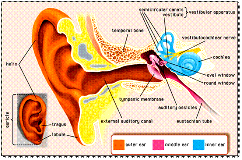
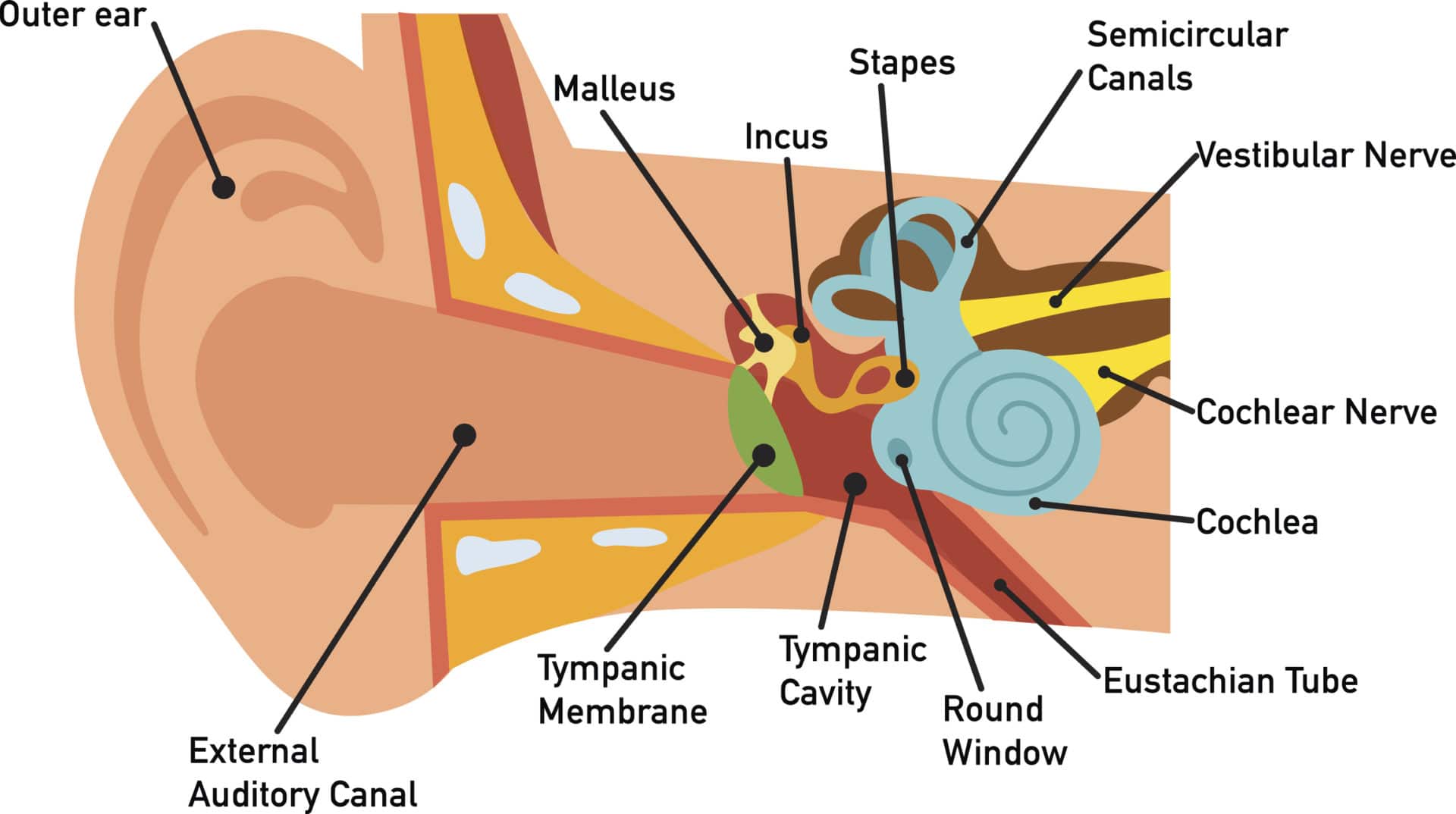
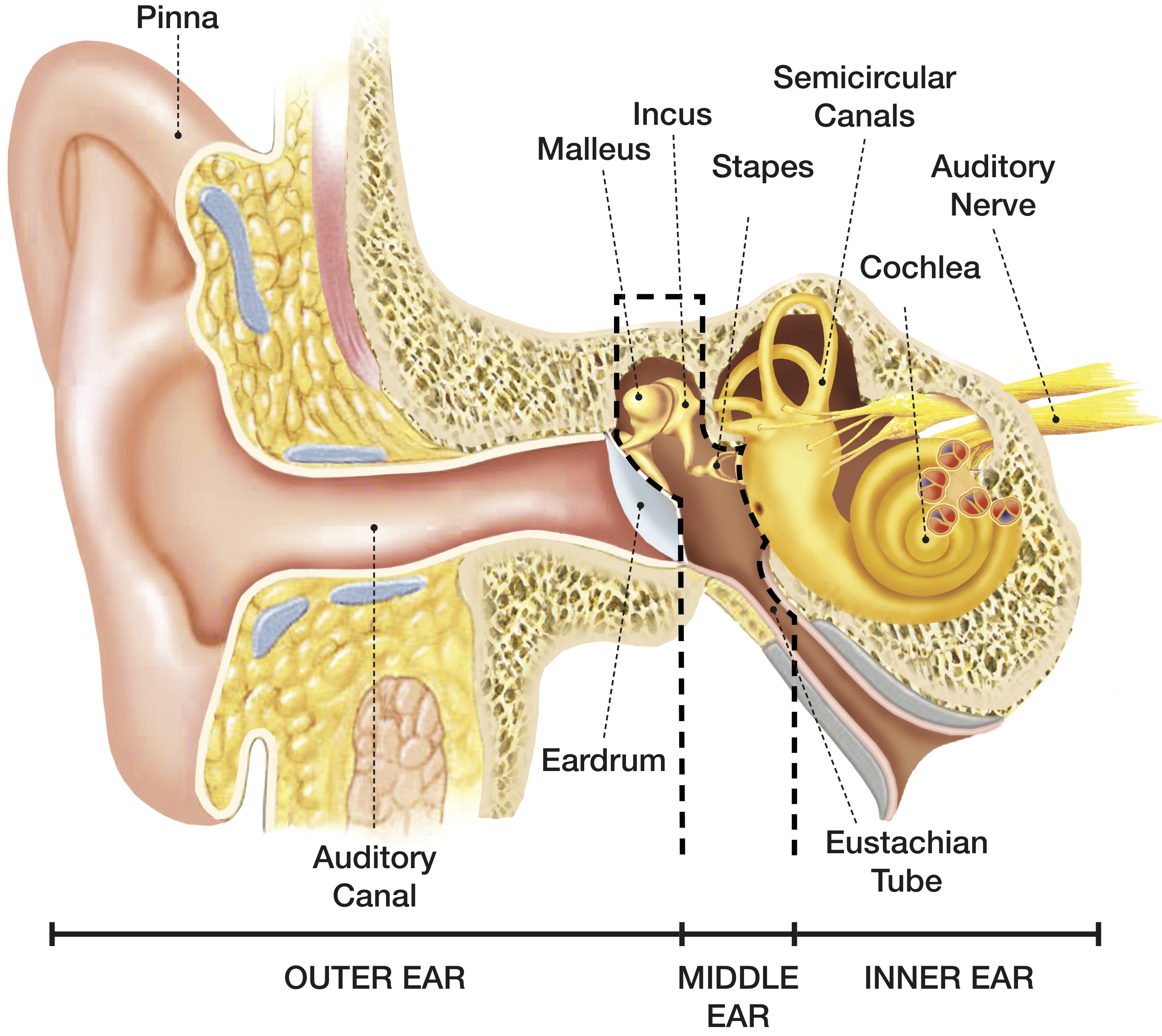
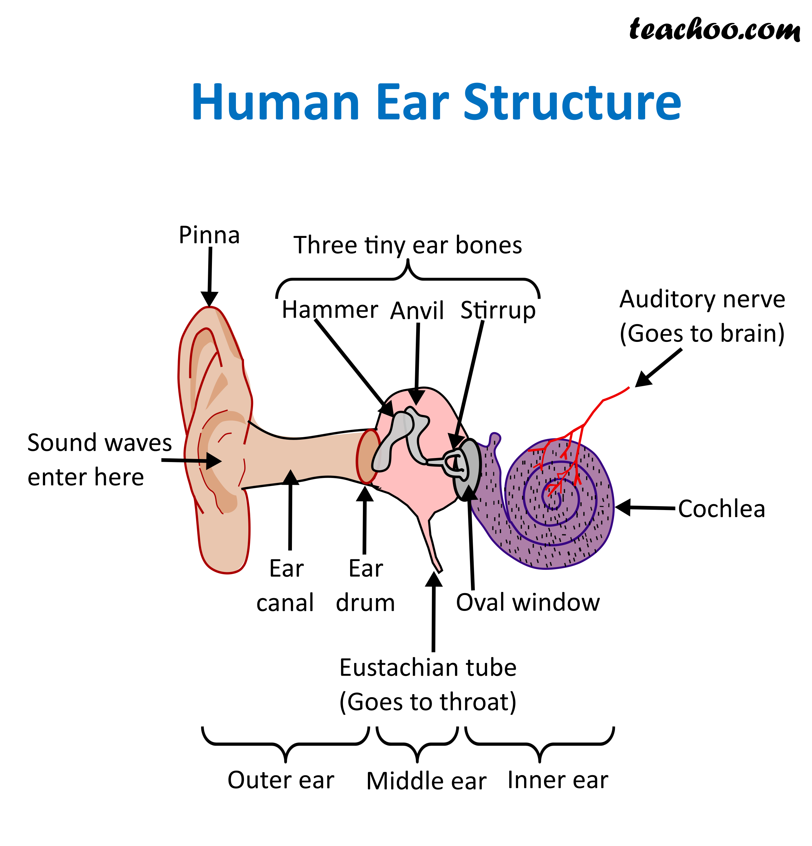
Labelled Diagram of Ear The Ear is composed of various parts which have been described below. Outer Ear: The outer Ear has two parts pinna or auricle and Ear canal Pinna or auricle: This part is present outside of the middle ear and pinna collects the sound waves and transmit them to the Ear cannal.
Outer Ear Anatomy Outer Ear Infection & Pain Causes & Treatment
Download a free printable outline of this video and draw along with us: https://artforall.me/video/how-to-draw-human-earThank you for watching. Please subsc.
Outer Ear Anatomy Outer Ear Infection & Pain Causes & Treatment
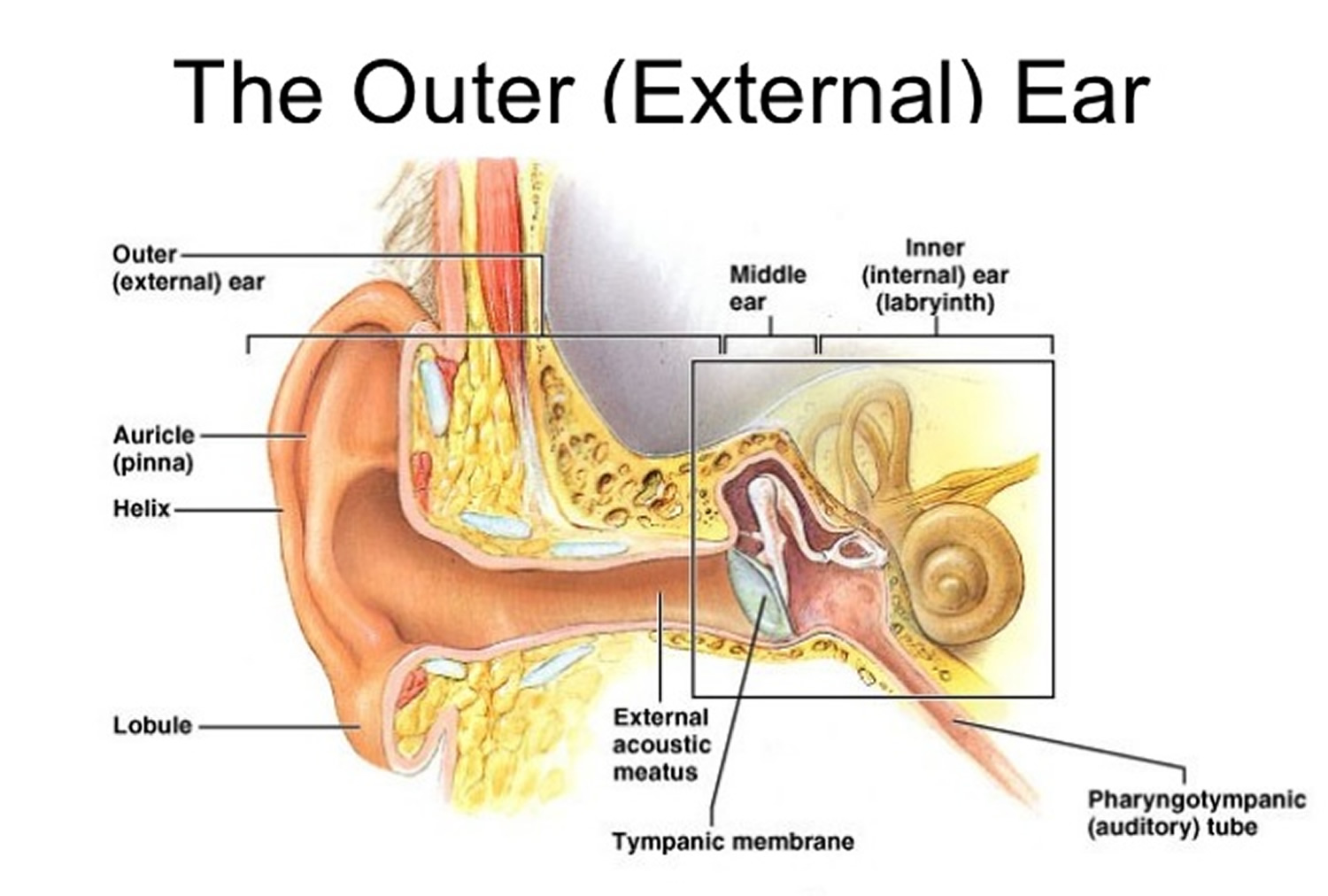
Human ear. The ear is divided into three anatomical regions: the external ear, the middle ear, and the internal ear (Figure 2). The external ear is the visible portion of the ear, and it collects and directs sound waves to the eardrum. The middle ear is a chamber located within the petrous portion of the temporal bone.
Understanding how the ear works Hearing Link Services
1: Diagram showing the structure of the human ear, detailing the parts. | Download Scientific Diagram Figure 2 - uploaded by Benjamin Gorman Content may be subject to copyright. 1:.
Human Ear Anatomy How Hearing Works Connect Hearing
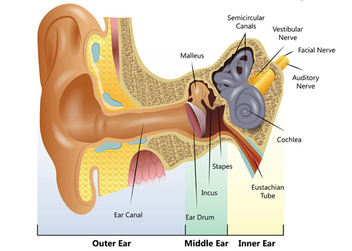
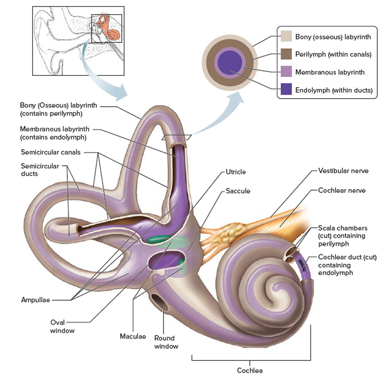
The ear also helps in balancing the body. The human ear allows us to feel the effect of gravity that is known as stationary balance and it also helps to feel the acceleration that is known as dynamic balance. The utricle and saccule provide a static balance. Dynamic balance is provided by semi-circular canals.
How You Hear Northland Audiology
Ear Anatomy - Inner Ear. Ear Anatomy Schematics. Ear Anatomy Images. Chapter 4 - Fluid in the ear. Fluid in the ear Discussion. Fluid in the ear Outline. Middle Ear Ventilation Tubes. Fluid in the ear Images. Chapter 5 - Traveler's Ear.

Human ear anatomy. Ears inner structure, organ of hearing ve (1000410) Infographics Design
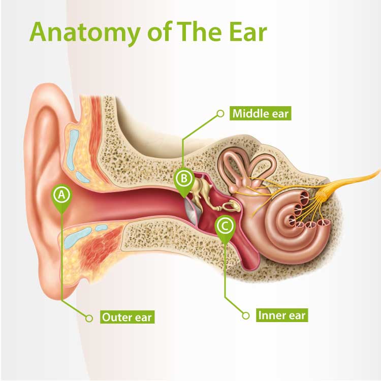
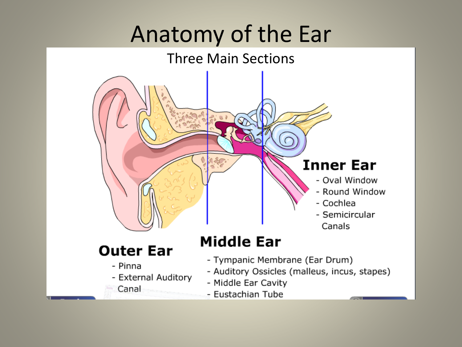
The ear diagram is one of the important topics for Class 10 and 12 students of the CBSE board and in this article, we will briefly explain the structure of the ear, its different parts and their functions. Parts of the Human Ear. The human ear consists of three different parts. These are: The outer ear. The middle ear. The inner ear

Associate Degree Nursing Physiology Review Ear anatomy, Middle ear, Human anatomy and physiology
Human Ear Diagram Wondering what is the structure of the human ear, and how it performs the function of hearing? Look no further, this Bodytomy article gives you a labeled human ear diagram and also explains the functions of its different components. Home / Uncategorized / Human Ear Diagram
3 Things to Know About the Signal Path of the Auditory System — Pro Audio Files
Thanks for watching our Channel. how to draw internal structure of human ear,diagram of human ear for class 8,diagram of human ear with labelling,structure o.
Anatomy of the Ear
human ear, organ of hearing and equilibrium that detects and analyzes sound by transduction (or the conversion of sound waves into electrochemical impulses) and maintains the sense of balance (equilibrium). Understand the science of hearing and how humans and other mammals perceive sound How humans and other mammals perceive sound.
How We Hear Hearing Associates, Inc.
Diagram of Ear Human ear is a sense organ responsible for hearing and body balance. The outer ear receives the sound waves and transmits them down the ear canal to the eardrum. This causes the eardrum to vibrate and sound is produced. The diagram of the ear is important from Class 10 and 12 perspectives and is usually asked in the examinations.

How The Ear Works
Overview Your outer ear and middle ear are separated by your eardrum, and your inner ear houses the cochlea, vestibular nerve and semicircular canals (fluid-filled spaces involved in balance and hearing). What is the ear? Your ears are organs that detect and analyze sound. Located on each side of your head, they help with hearing and balance.

Ears and hearing How do they work?
1/4 Synonyms: External auditory meatus, External acoustic pore , show more. The ear is a complex part of an even more complex sensory system. It is situated bilaterally on the human skull, at the same level as the nose. The main functions of the ear are, of course, hearing, as well as constantly maintaining balance.
Human Ear Anatomy Parts of Ear Structure, Diagram and Ear Problems
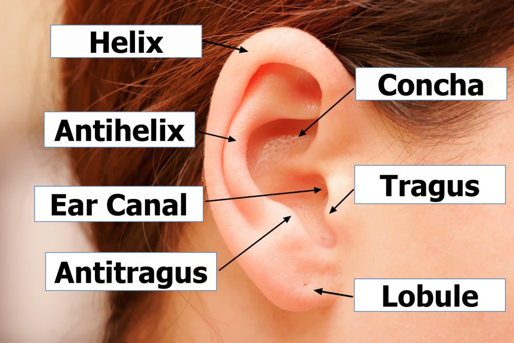
1. External Ear: It comprises a pinna, external auditory meatus (canal) & tympanic membrane. (i) Pinna: ADVERTISEMENTS: The pinna is a projecting elastic cartilage covered with skin. Its most prominent outer ridge is called the helix.

Labeled Diagram Of The Interior Of A Human Ear For Reference stock vector art 185868456 iStock
A labelled cross-sectional diagram of the human ear. Summary [ edit] All translations [ edit] Translations added to this section should be free of copyright claims (either CC0 or public domain). Malleus ≅ malleus (Q319636) Malleus Incus ≅ incus (Q497385) Incus Stapes ≅ stapes (Q499897) Stapes oval window ≅ oval window (Q2301793) oval window
Structure and Function of Human Ear with Diagram Teachoo
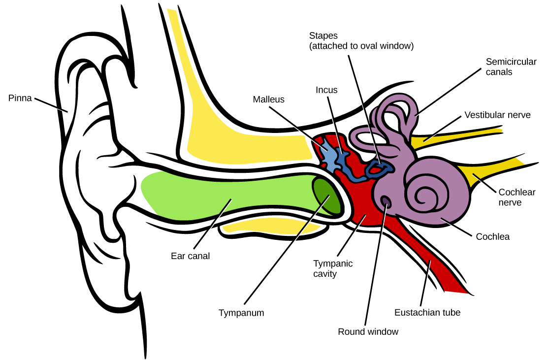
The Human Ear www.TurnItToTheLeft.com. If the noise is too loud, walk away, turn it down (Turn it to the Left), or use ear plugs. pinna ear canal ear drum hammer anvil stirrup Eustachian tube (connects to the nose) cochlea semicircular canals nerves (connect to the brain) Directions: Color in the diagram below using a different color for.
